Patient specific 3D printed implants

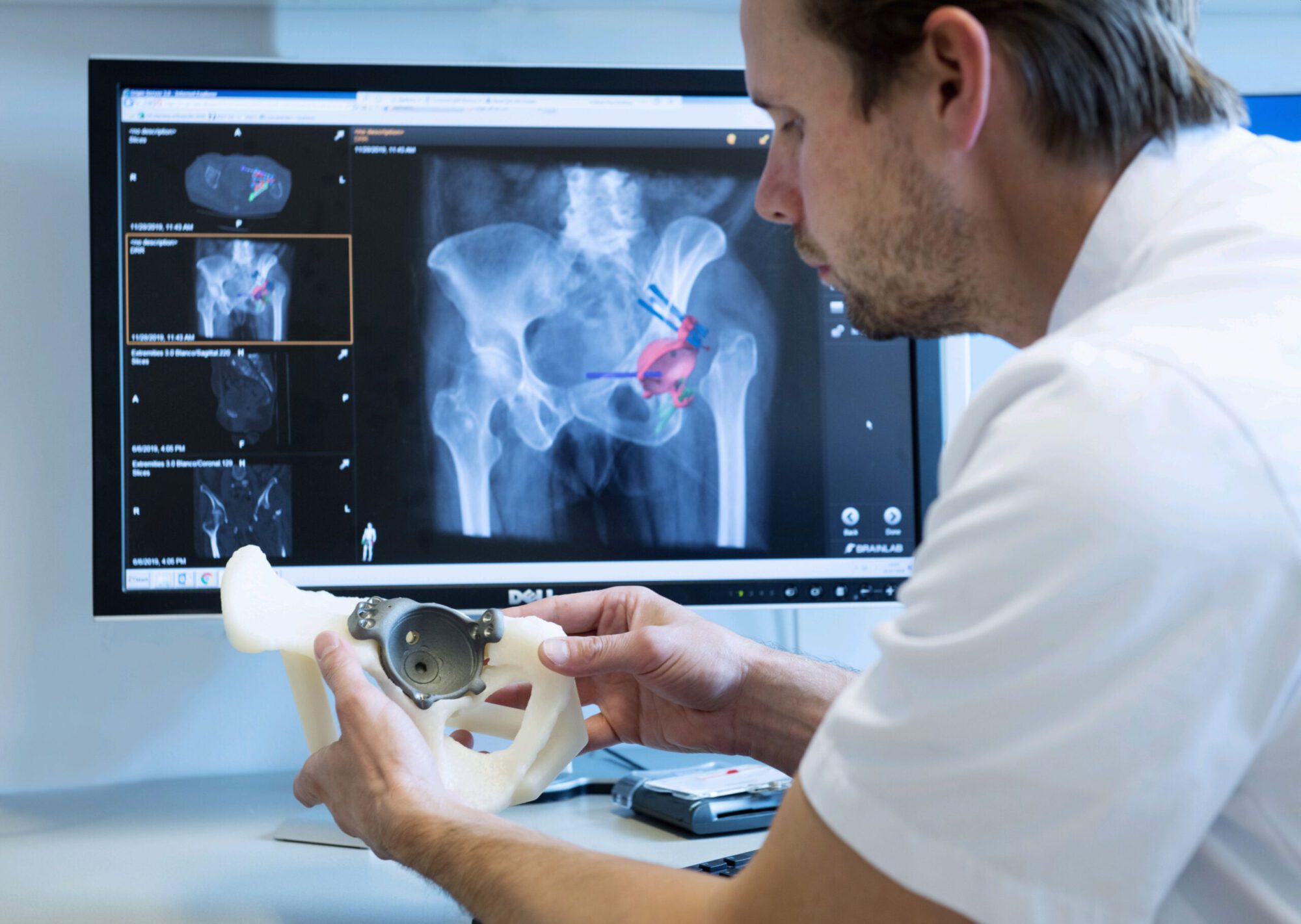
Custom designed 3D printed implants offer a solution in patients where other reconstruction options are not available. For the design and fabrication of these implants, we collaborate with different companies.
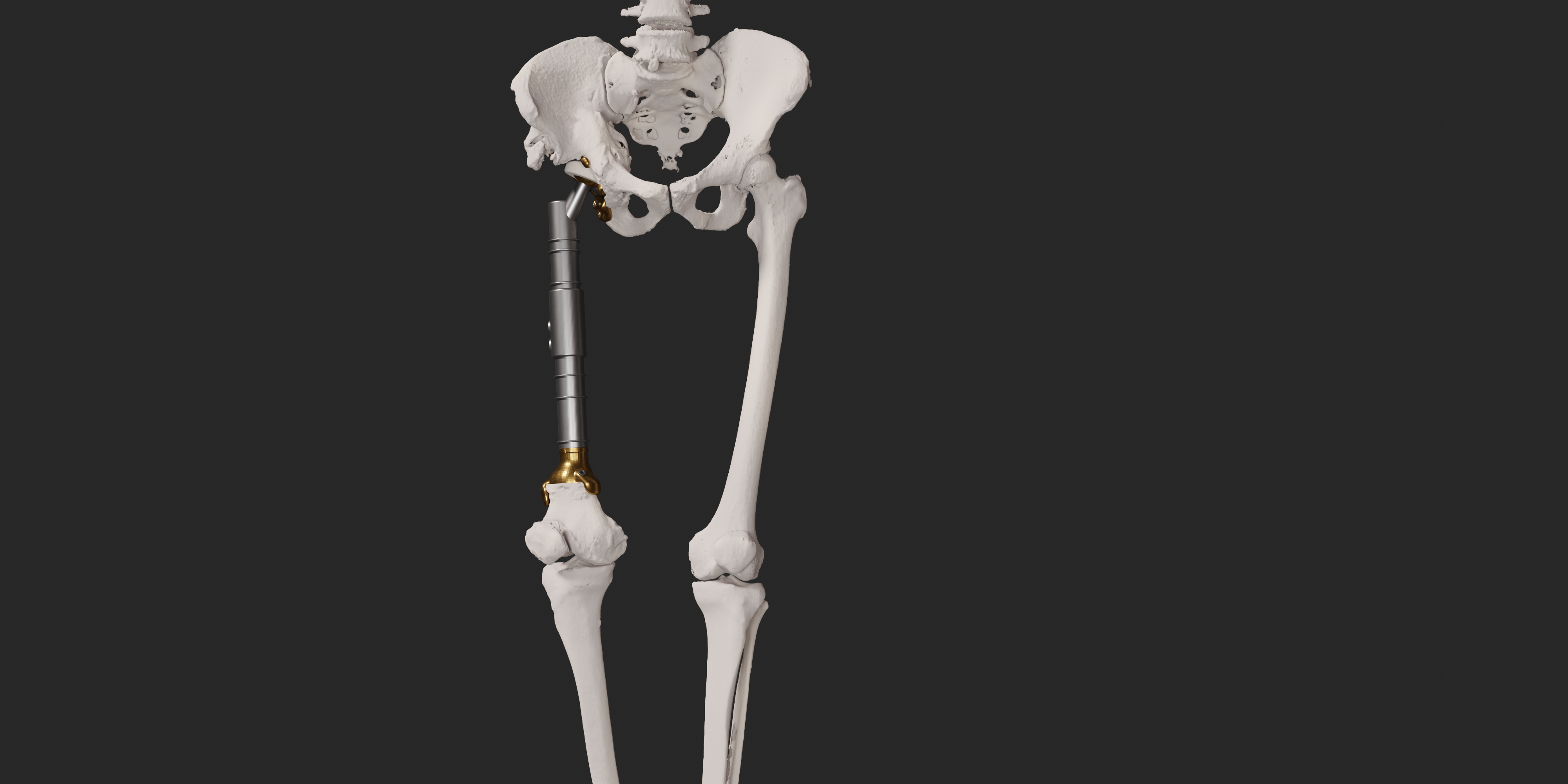
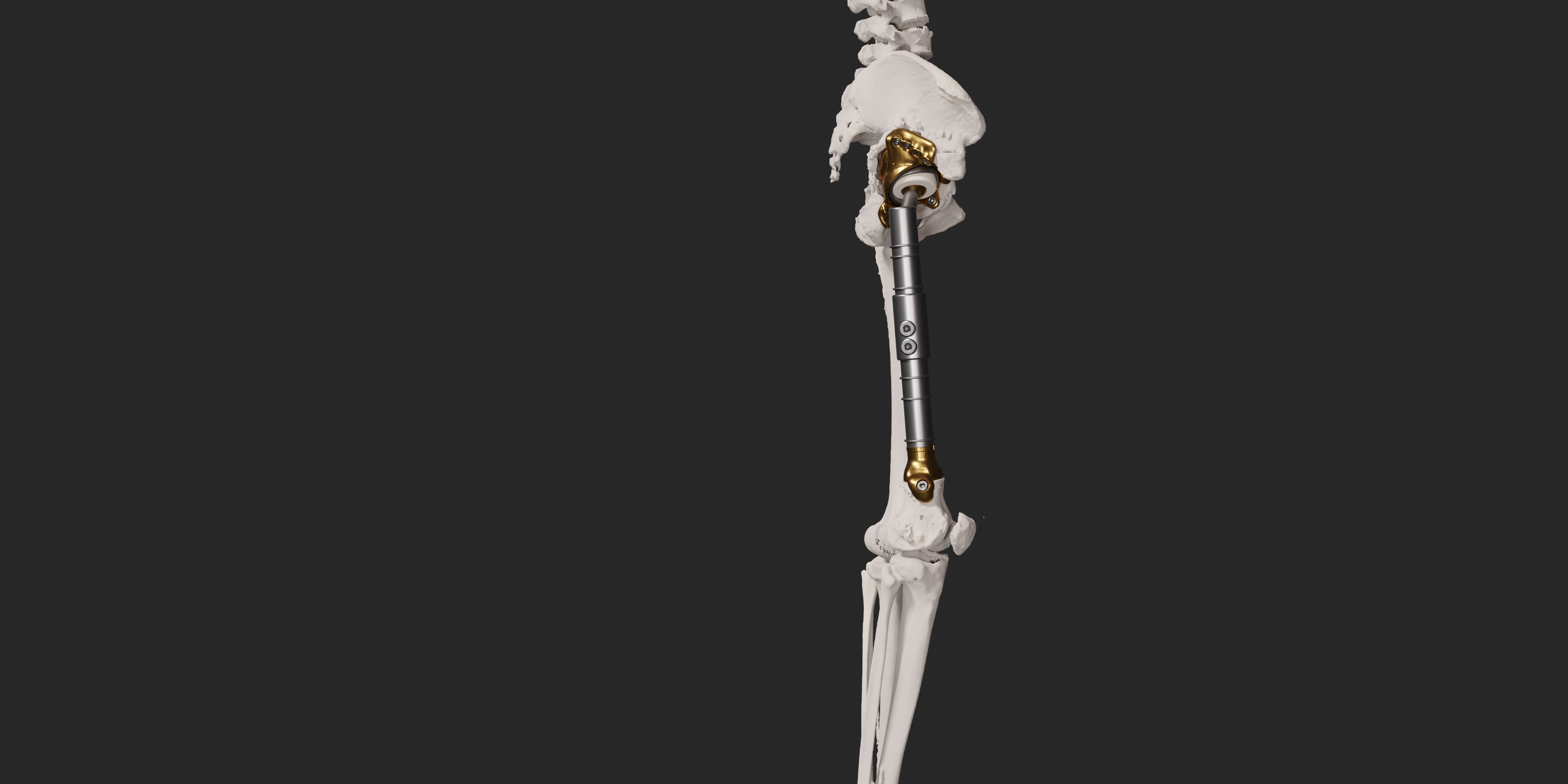
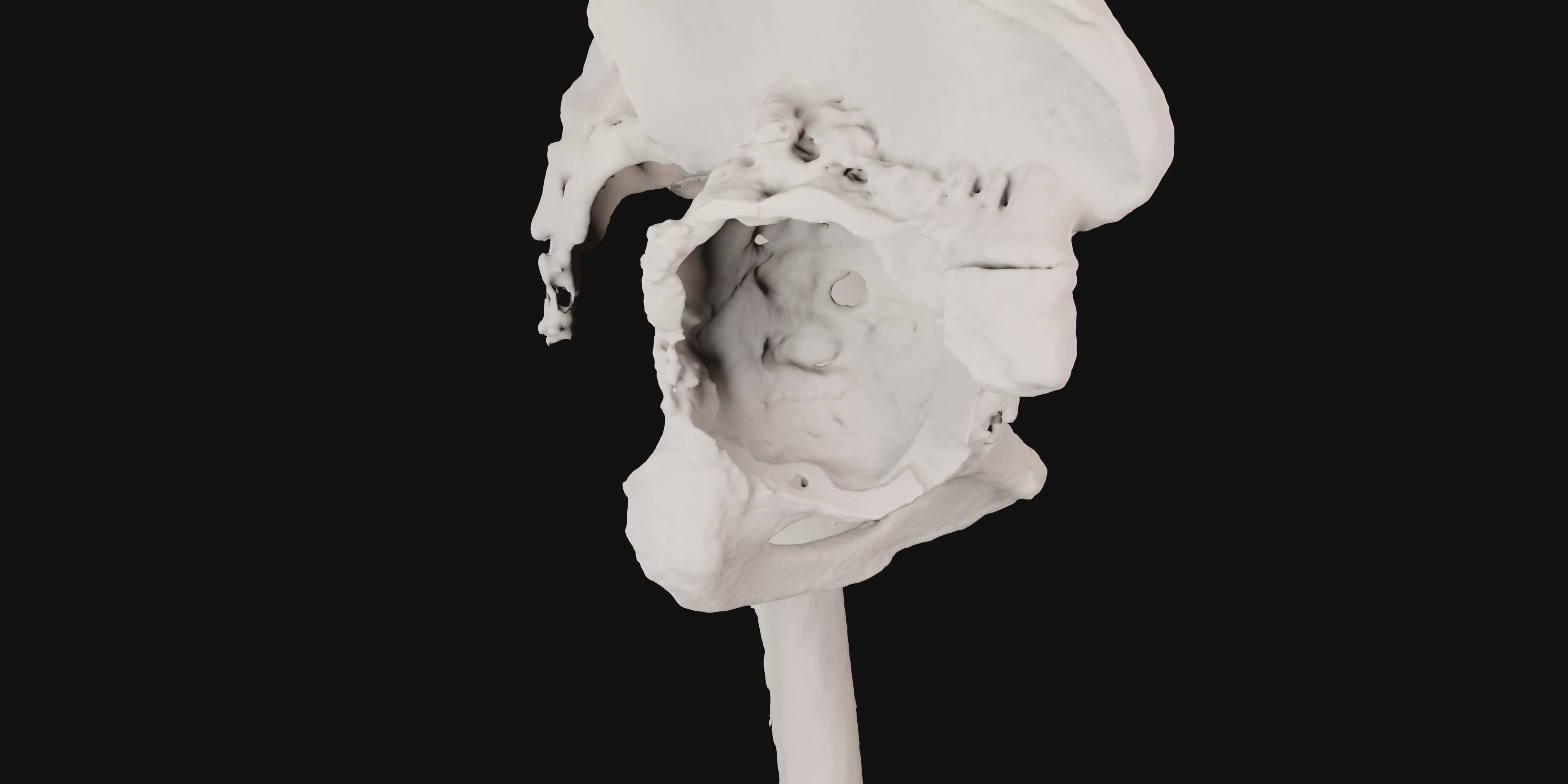
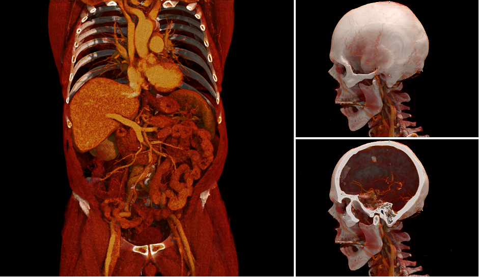
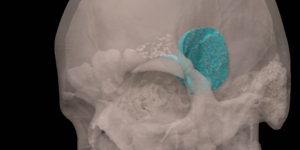
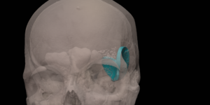
A 41 year old female suffered a combination of a large acetabular and a subtotal femoral defect, due to a chronic low grade periprosthetic joint infection of a revision total hip replacement.
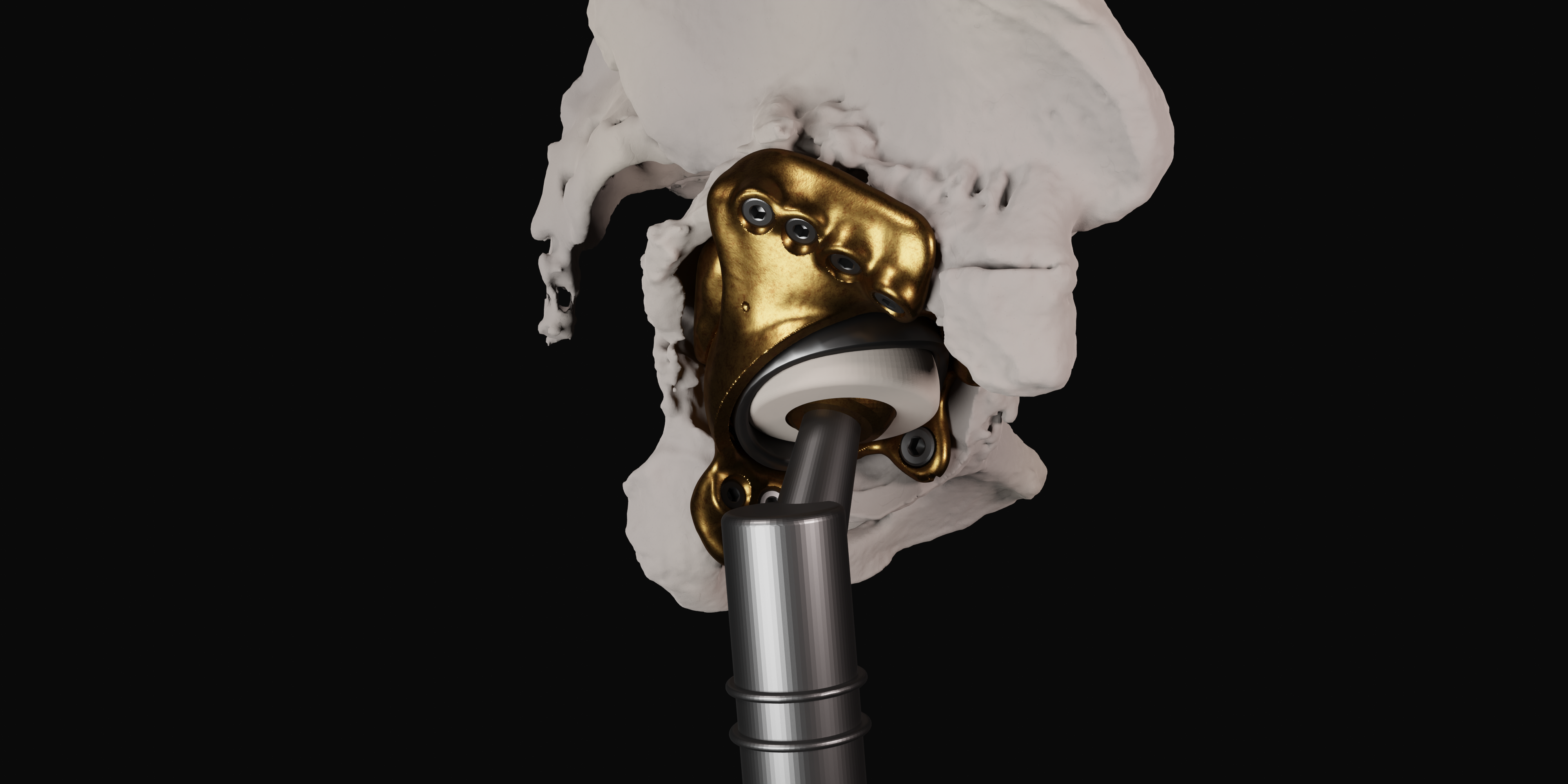
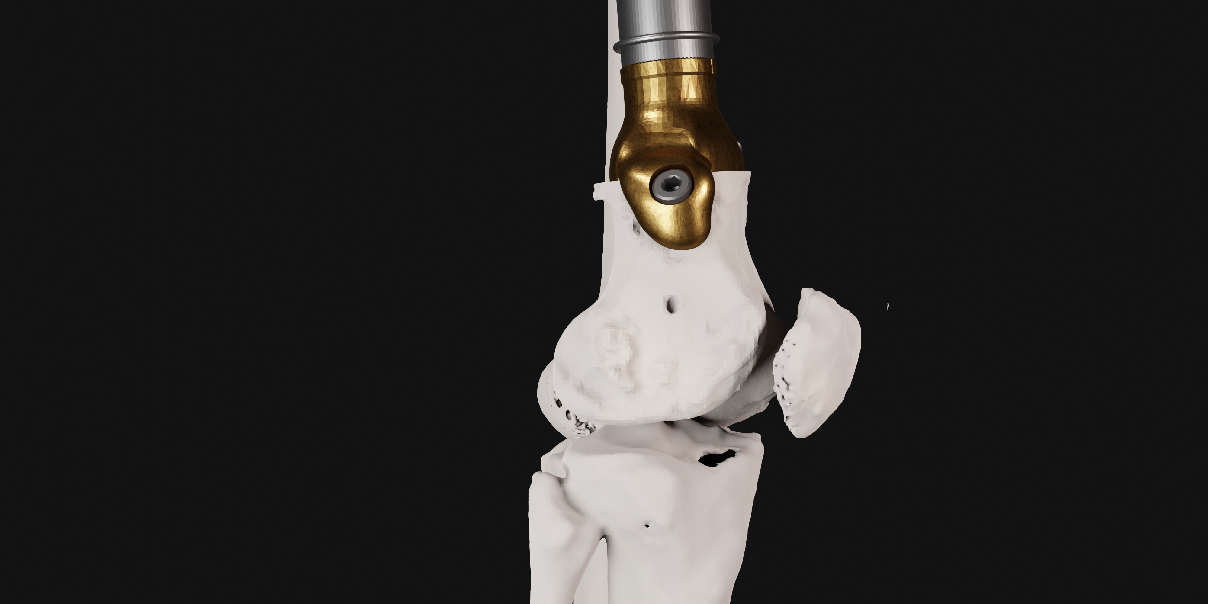
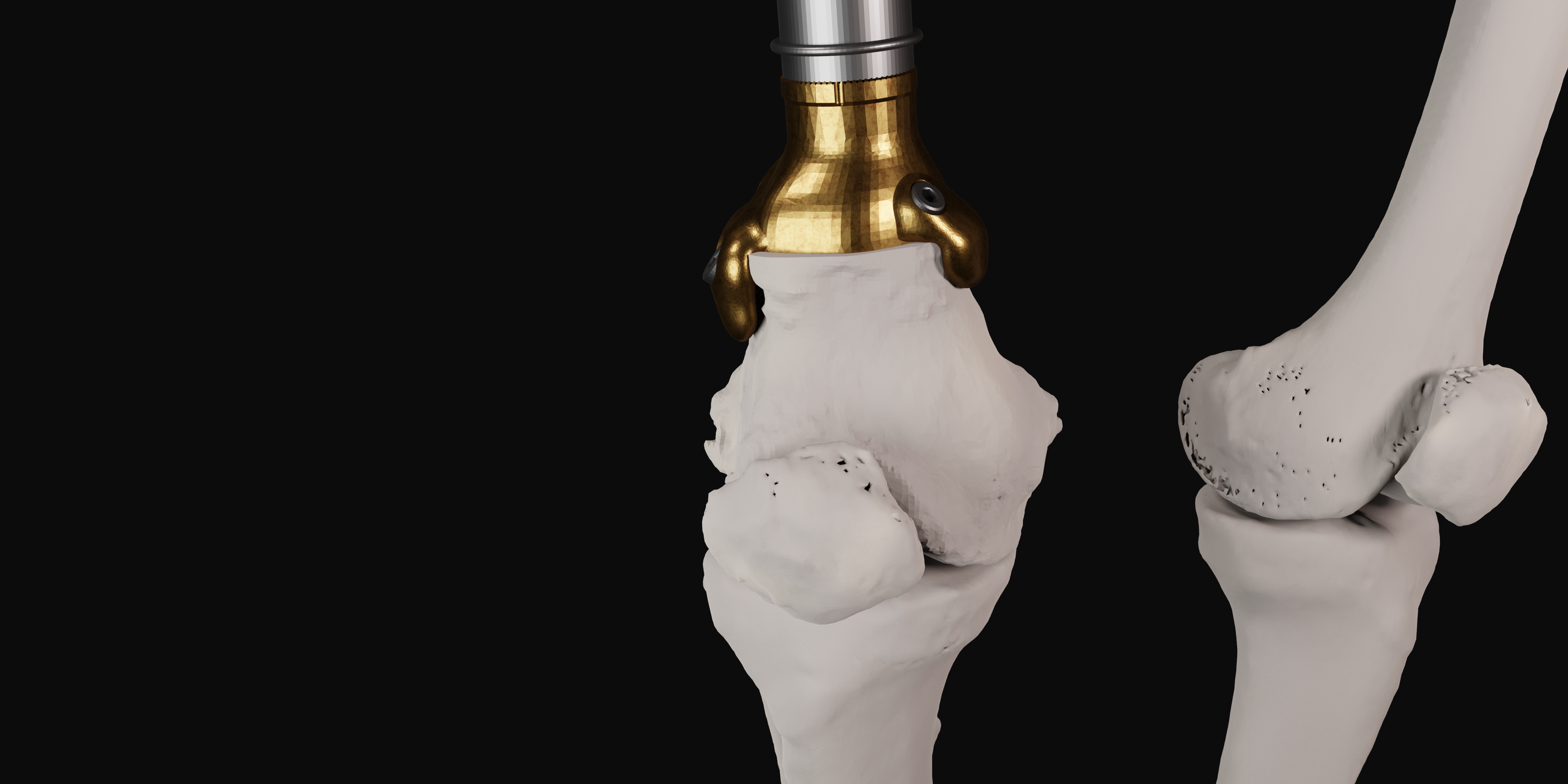
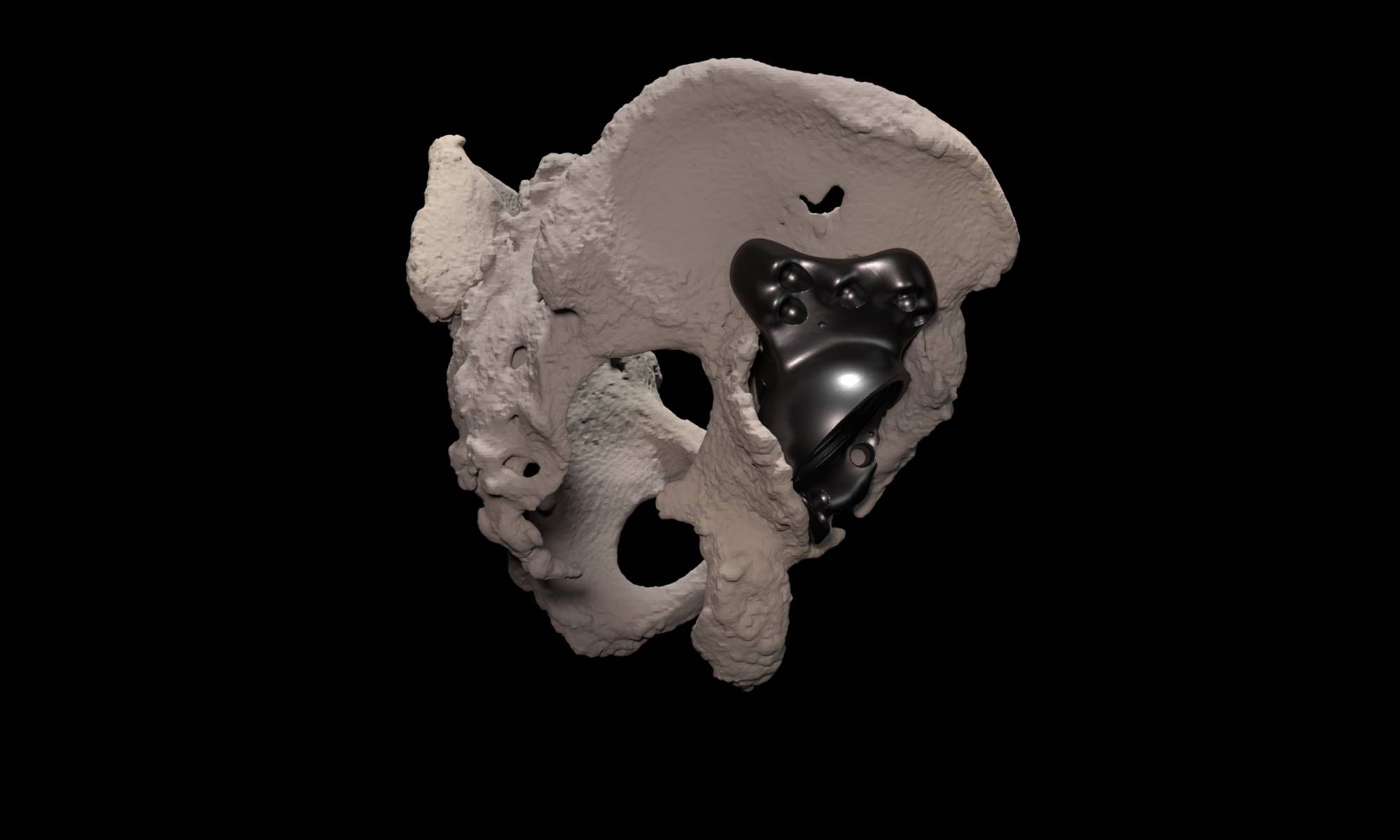
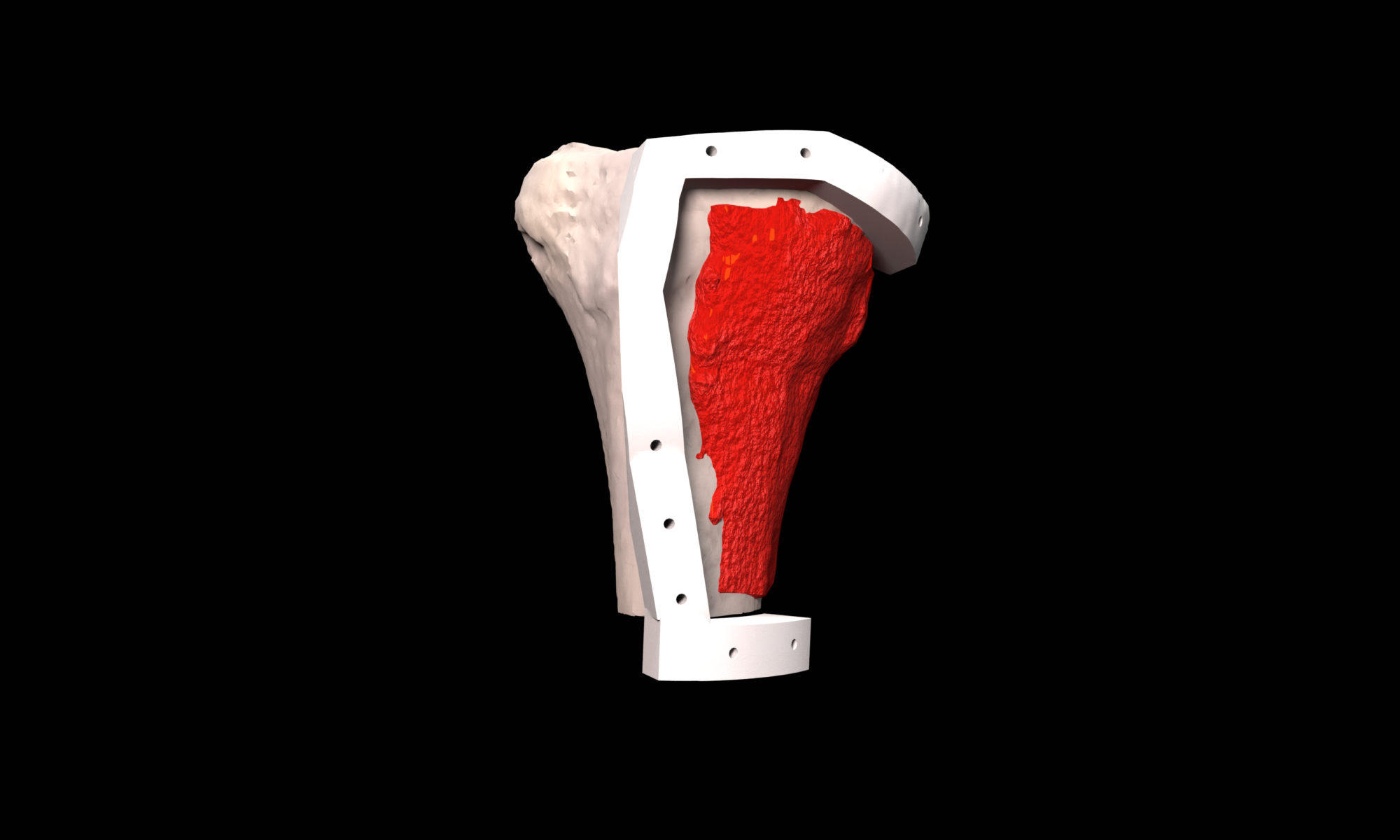
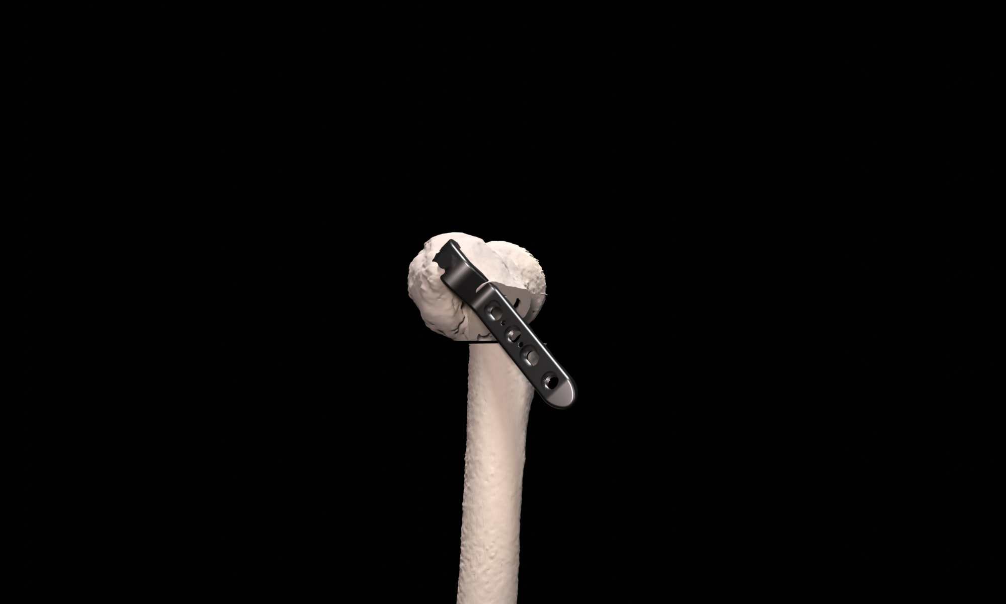
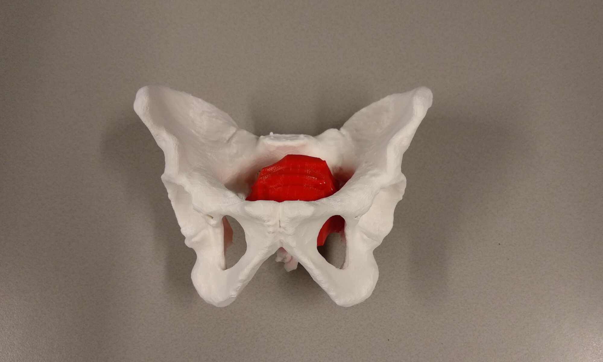
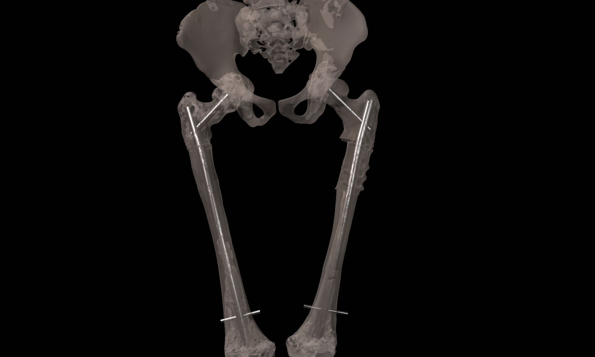
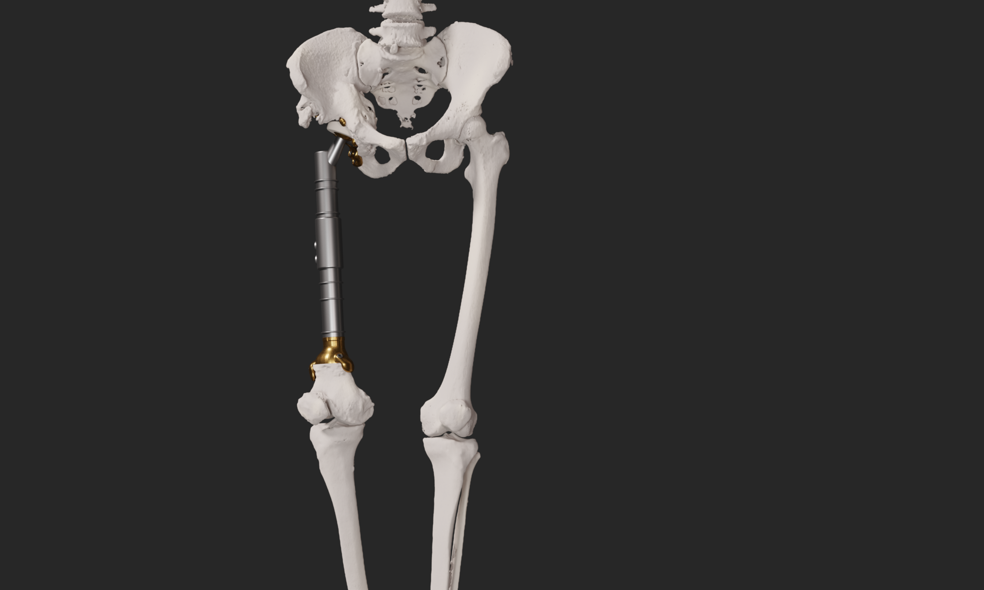
The final procedure included reconstruction of the pelvis with an OSSISĀ® AceOs plus custom acetabular implant. Femoral reconstruction was performed using a MUTARSĀ® proximal femoral replacement with a custom MUTARS C-fit 3D printed joint sparing fixation, preserving the native knee joint and with this preventing the necessity for total femoral replacement.

Patient specific 3D printed hip implants

Patient specific 3D printed hip implants offer a novel method to reconstruct large pelvic defects. Patients with such defects are in general not ambulatory and have large disabilities. These custom designed implants offer a solution in patients where other reconstruction options are not available. For the design and fabrication of these implants, we collaborate with OSSIS, a New Zealand based company.

A multi center study is on going to study the fixation of these implants using Radiostereometry.

Imaging Services Group

The Imaging Services Group (ISG) at the department of Radiology, Leiden University Medical Center, is a specialized image processing group responsible for extracting relevant information from medical images for diagnosis and image-guided treatment decisions. This includes virtual surgical planning for liver resections, radiological reporting for oncological follow-up, and creation of 3D reconstructions for orthopaedic cases. The ISG is responsible for delivering high quality information to radiologists by using state-of-the art image processing software (including AI) that can be used for radiological reporting in clinical routine. Furthermore, the ISG is involved in many different research projects focusing on the development and introduction of new innovations in the field of imaging, as well as determining their added value in clinical practice.

Allograft Reconstruction

Biological reconstruction is still the preferred method to reconstruct bone defects caused by bone tumor resection in young patients. The removal of the tumor including a required margin is 3D virtually planned in which the information of different imaging modalities, such as CT, MRI and PET, is combined. The surgical resection plan is translated to the operation room by designing patient specific surgical guides that uniquely fit the patient anatomy and indicate the virtually planned resection. To validate the use of these guides surgical navigation is used during surgery. Data from a digital bonebank, that contains 3D models of all available allograft bones, is used to select the best matching allograft bone and to design resection guides for accurate allograft preparation during surgery.

Proximal Femur Osteotomy

Complex proximal femur deformities can result after (pathological) fracture mal-union. Deformities in this location can be very invalidating due to the relation to the hip joint and negative effect on leg-length, rotations and limb biomechanics. Proximal femoral deformities can be treated by corrective osteotomies and are preferably secured with a compressive blade plate. In complex cases, careful surgery planning in 3D is necessary to achieve the desired correction in multiple planes. A key part of this procedure is to position the blade of the plate in the femoral head and neck correctly. First, the desired post-operative correction and blade position is virtually planned using 3D models generated from 3D CT scans. Second, to translate the virtual planning to the patient in the operating room, patient specific guides are designed, 3D printed and sterilized for intra-operative use. These custom surgical guides, that precisely fit the unique bony shape of the patient, indicate the desired osteotomy location, osteotomy planes and blade direction and facilitate the execution surgical procedure with high accuracy.

Anatomical Models

For surgical preparation and shared decision making between multidisciplinary surgical teams, patient specific, multi colored 3D printed anatomical models can be very useful. 3D models of (bony) pathology also have a high educational value for surgeons in training. Also paramount is the use of 3D printed anatomical models for patient insight into their disease and shared decision making between surgeon and patient.

Trauma

After trauma a part of the skull might be damaged or lost. A reconstruction of the skull can be performed using patient specific implants. The implants are designed to exactly fit the defect and restore the patient anatomy. Reconstruction includes the skull bone, but can also incorporate surrounding structures such as the orbital roof, orbital wall, orbital rim or zygoma. The patient specific 3D virtually designed implants enables us to perform reconstructions that may have not been possible using conventional reconstruction methods.

Oncology

When a tumor invades the skull bone and surgical removal is indicated, the procedure is 3D virtually planned. The removal of the tumor including a required margin is 3D virtually planned combining information from different imaging modalities, such as CT and MRI. The surgical resection planning is translated to the operation room by designing patient specific surgical guides that uniquely fit the patient anatomy and indicate the virtually planned resection during surgery. Because the use of a surgical cutting guide creates a predictable bone defect, a patient specific skull implant can be designed to accurately reconstruct the bone defect.

Spheno-Orbital Meningioma

In the LUMC, a specialized multidisciplinary team treats patients with meningiomas. For patients with a spheno-orbital meningioma, the resection and reconstruction is 3D virtually planned. The resection is performed using a patient specific resection guide that uniquely fits the patient anatomy. Subsequently, the bone defect is reconstructed using a patient-specific implant, reconstructing both the temporal bone and the lateral orbital wall, if indicated.

Full Femur Osteotomy

A Shepherd’s Crook deformity is caused by fibrous dysplasia and can be treated by a corrective osteotomy secured with eighter plates or intra-medullary nails. In complex cases, multiple and very large corrections are necessary, requiring careful surgical planning in 3D. First, the desired post-operative correction is virtually planned using 3D models generated from 3D CT scans. Second, to translate the virtual planning to the patient in the operating room, patient specific osteotomy guides are designed, 3D printed and sterilized for intra-operative use. These custom surgical guides, that precisely fit the unique bony shape of the patient, indicate the desired osteotomy location and osteotomy planes and facilitate the execution of the surgical procedure with high accuracy.

The patient in the example case had two surgeries to correct both legs.