Allograft Reconstruction

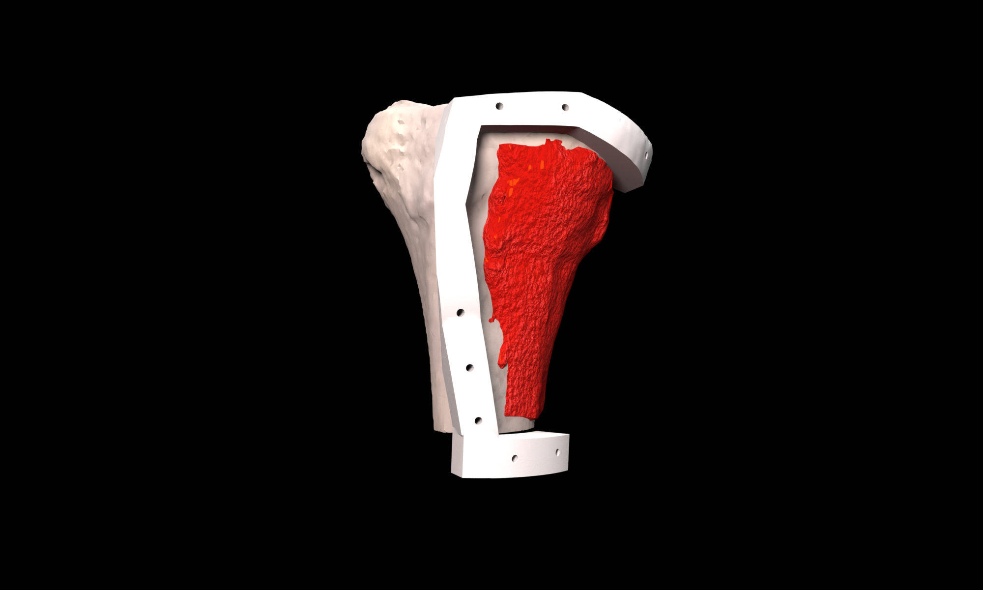
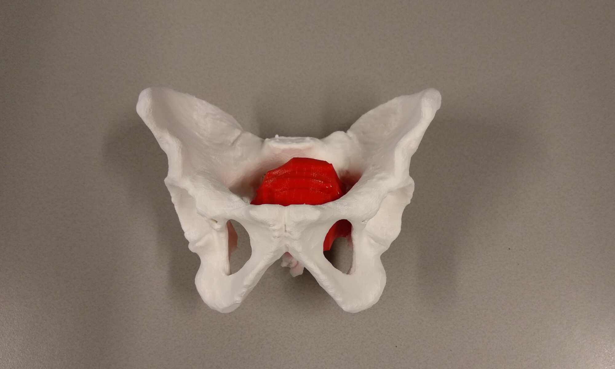
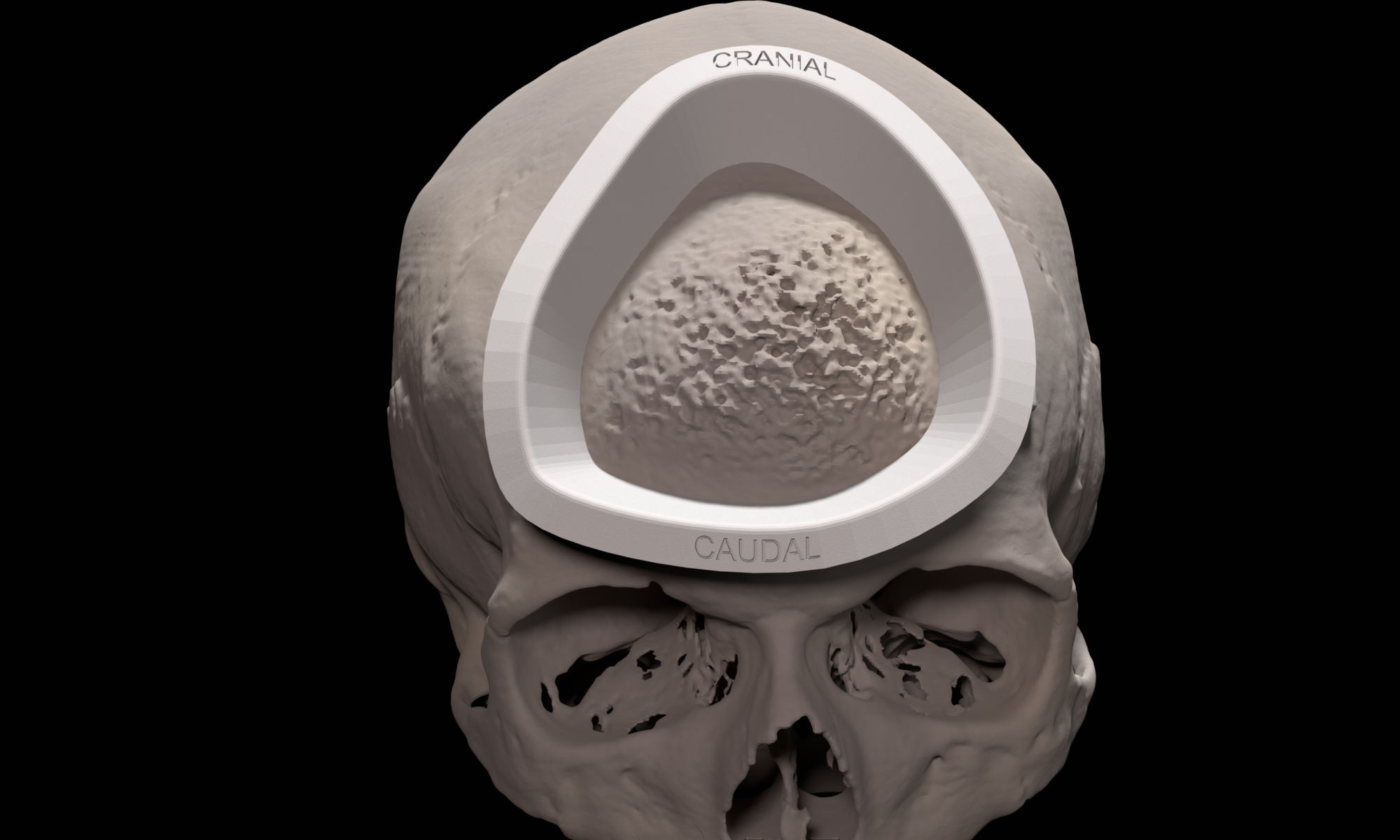
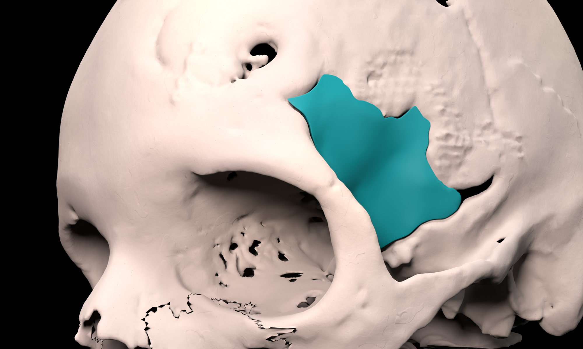
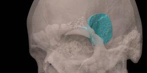
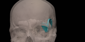
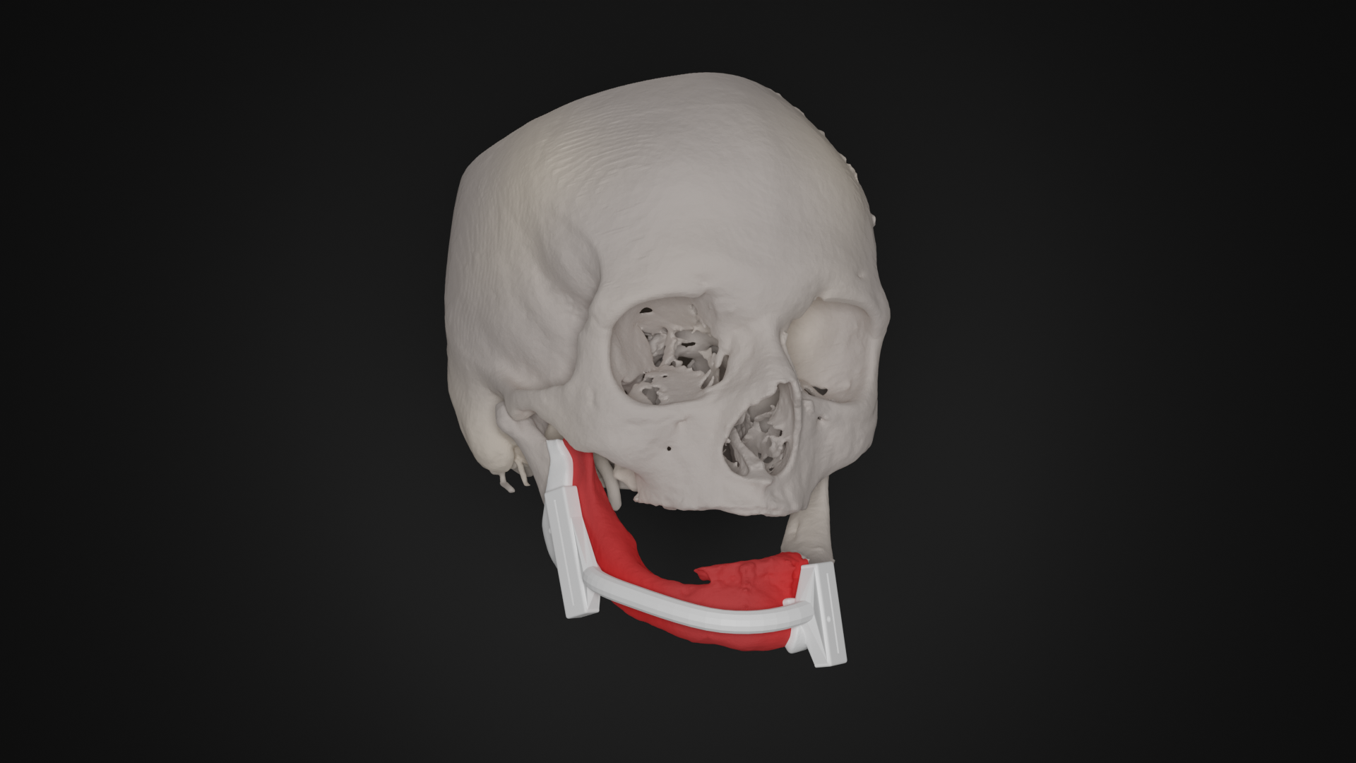
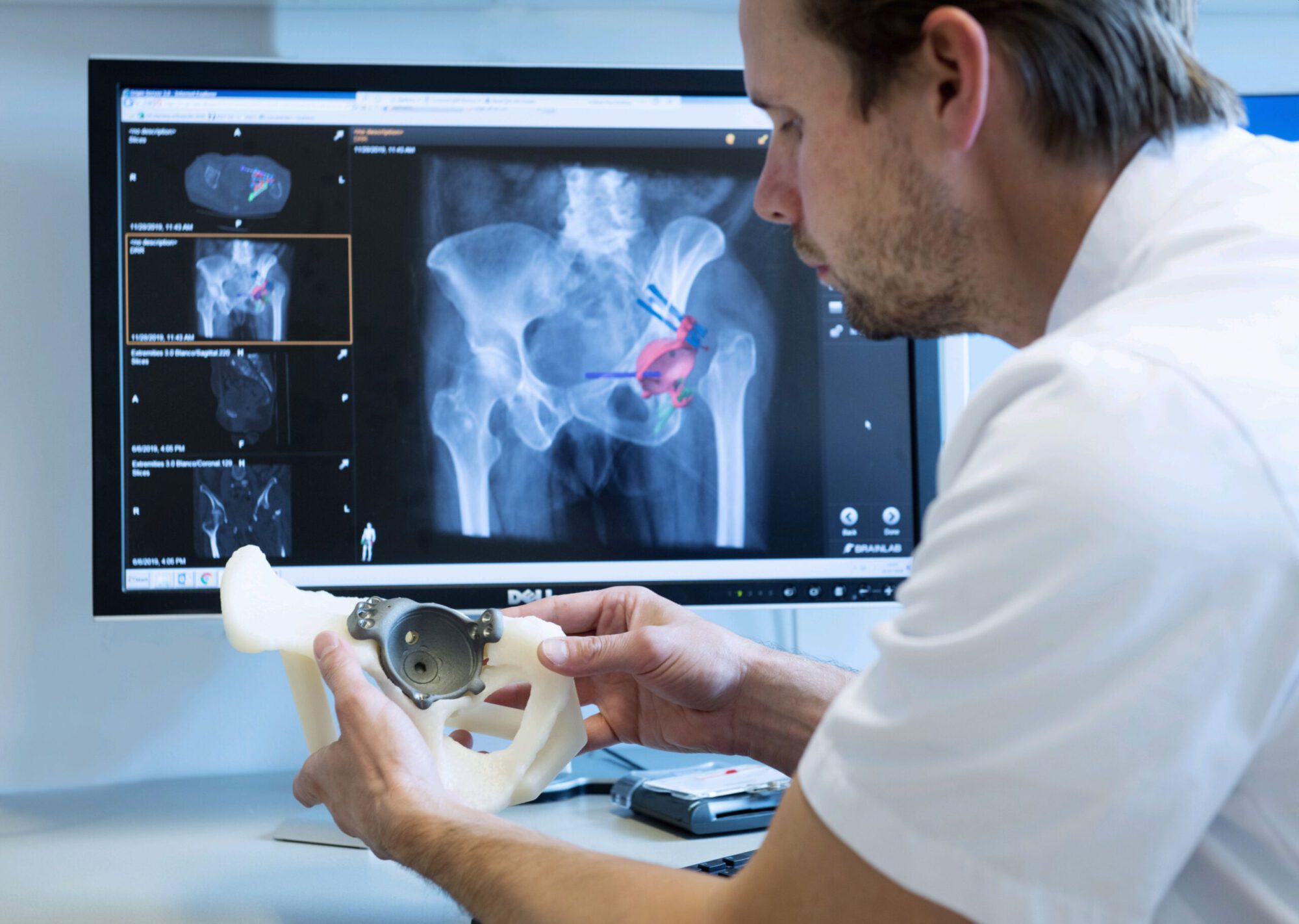
Biological reconstruction is still the preferred method to reconstruct bone defects caused by bone tumor resection in young patients. The removal of the tumor including a required margin is 3D virtually planned in which the information of different imaging modalities, such as CT, MRI and PET, is combined. The surgical resection plan is translated to the operation room by designing patient specific surgical guides that uniquely fit the patient anatomy and indicate the virtually planned resection. To validate the use of these guides surgical navigation is used during surgery. Data from a digital bonebank, that contains 3D models of all available allograft bones, is used to select the best matching allograft bone and to design resection guides for accurate allograft preparation during surgery.
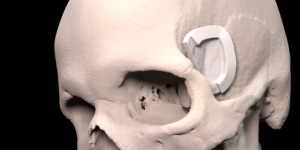
Anatomical Models

For surgical preparation and shared decision making between multidisciplinary surgical teams, patient specific, multi colored 3D printed anatomical models can be very useful. 3D models of (bony) pathology also have a high educational value for surgeons in training. Also paramount is the use of 3D printed anatomical models for patient insight into their disease and shared decision making between surgeon and patient.

Oncology

When a tumor invades the skull bone and surgical removal is indicated, the procedure is 3D virtually planned. The removal of the tumor including a required margin is 3D virtually planned combining information from different imaging modalities, such as CT and MRI. The surgical resection planning is translated to the operation room by designing patient specific surgical guides that uniquely fit the patient anatomy and indicate the virtually planned resection during surgery. Because the use of a surgical cutting guide creates a predictable bone defect, a patient specific skull implant can be designed to accurately reconstruct the bone defect.

Spheno-Orbital Meningioma

In the LUMC, a specialized multidisciplinary team treats patients with meningiomas. For patients with a spheno-orbital meningioma, the resection and reconstruction is 3D virtually planned. The resection is performed using a patient specific resection guide that uniquely fits the patient anatomy. Subsequently, the bone defect is reconstructed using a patient-specific implant, reconstructing both the temporal bone and the lateral orbital wall, if indicated.

Oncology

Removal of a tumor in the oral cavity that has invaded into the bone may require the removal of a part of the bone, thereby losing continuity of the bone. The removal of the tumor including a required margin is 3D virtually planned in which the information of different imaging modalities is combined, such as CT and MRI. The surgical resection planning is translated to the operation room by designing patient specific surgical guides that uniquely fit the patient anatomy and indicate the virtually planned resection during surgery. Using these same techniques, a reconstruction of the bone using fibula bone can be 3D virtually planned and transferred to the operating room using a patient specific reconstruction guide.