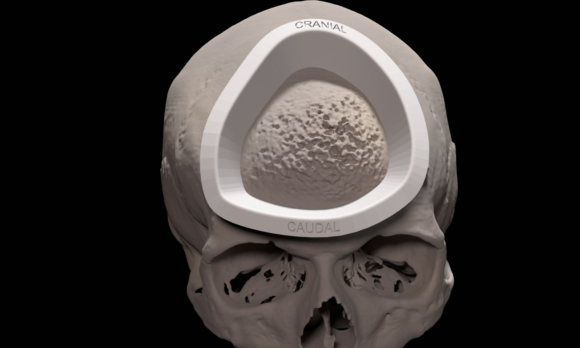
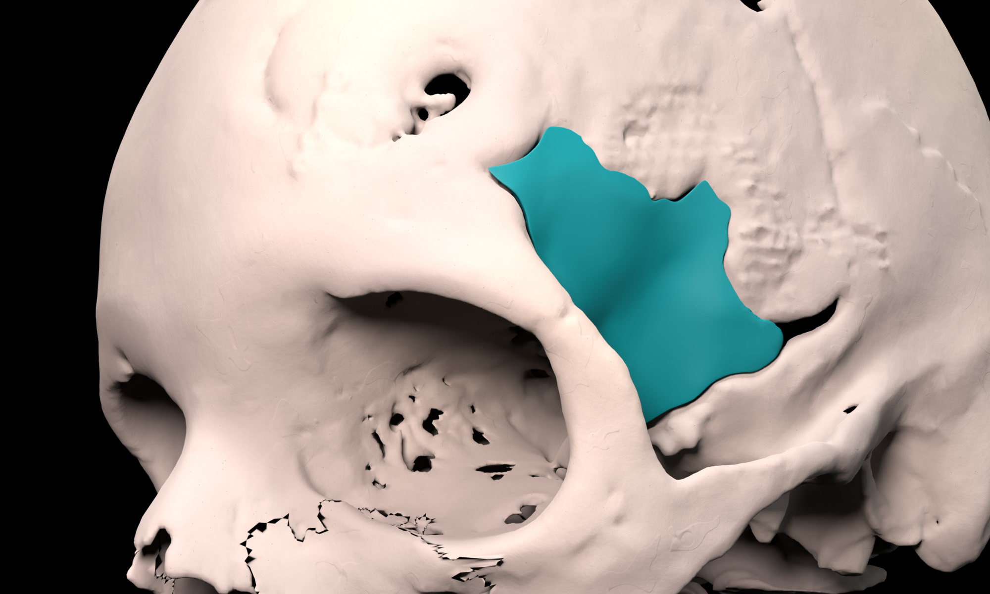
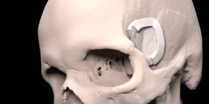
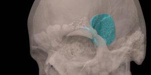
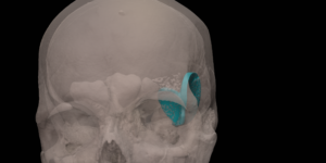
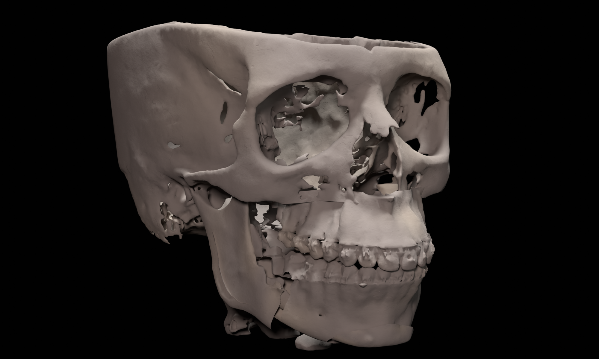
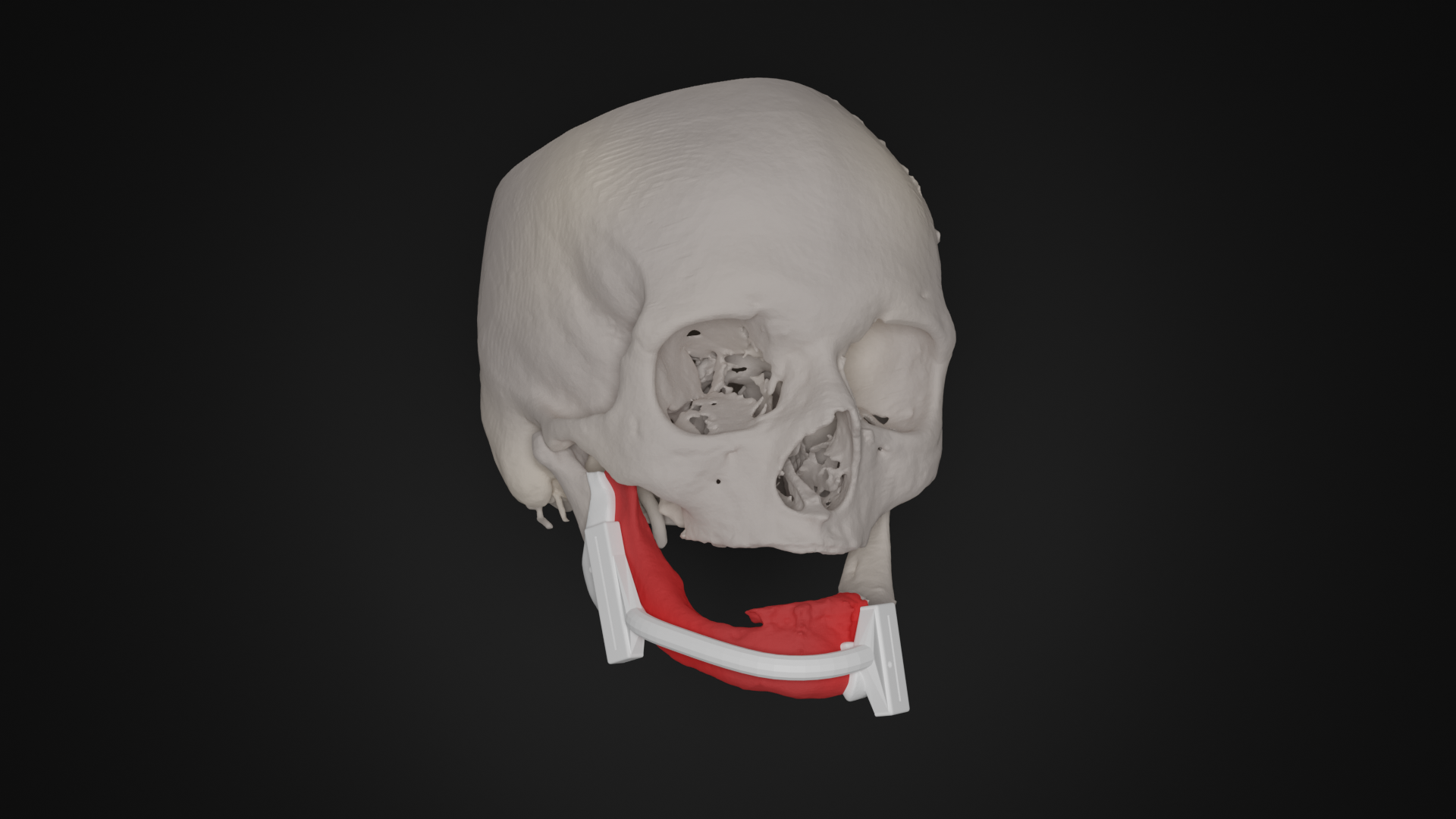
Spheno-Orbital Meningioma

In the LUMC, a specialized multidisciplinary team treats patients with meningiomas. For patients with a spheno-orbital meningioma, the resection and reconstruction is 3D virtually planned. The resection is performed using a patient specific resection guide that uniquely fits the patient anatomy. Subsequently, the bone defect is reconstructed using a patient-specific implant, reconstructing both the temporal bone and the lateral orbital wall, if indicated.

Full Femur Osteotomy

A Shepherd’s Crook deformity is caused by fibrous dysplasia and can be treated by a corrective osteotomy secured with eighter plates or intra-medullary nails. In complex cases, multiple and very large corrections are necessary, requiring careful surgical planning in 3D. First, the desired post-operative correction is virtually planned using 3D models generated from 3D CT scans. Second, to translate the virtual planning to the patient in the operating room, patient specific osteotomy guides are designed, 3D printed and sterilized for intra-operative use. These custom surgical guides, that precisely fit the unique bony shape of the patient, indicate the desired osteotomy location and osteotomy planes and facilitate the execution of the surgical procedure with high accuracy.

The patient in the example case had two surgeries to correct both legs.

Trauma

For trauma cases, 3D models in which mobile bone segments have been repositioned according to the normal patient anatomy are 3D printed and used to pre-bend reconstruction plates, saving time during surgery.

Orthognathic surgery

Orthognathic surgery involves a correction of the position of the lower and/or upper jaw. The desired post-operative position of the lower and/or upper jaw are virtually planned using 3D CT scans, clinical measurements and photographs. To translate the 3D virtual planning to the patient in the operating room, patient specific surgical guides are designed and 3D printed. These surgical guides that fit the dentition of the patient indicate the desired translation and rotation of the jaws with high accuracy.

Oncology

Removal of a tumor in the oral cavity that has invaded into the bone may require the removal of a part of the bone, thereby losing continuity of the bone. The removal of the tumor including a required margin is 3D virtually planned in which the information of different imaging modalities is combined, such as CT and MRI. The surgical resection planning is translated to the operation room by designing patient specific surgical guides that uniquely fit the patient anatomy and indicate the virtually planned resection during surgery. Using these same techniques, a reconstruction of the bone using fibula bone can be 3D virtually planned and transferred to the operating room using a patient specific reconstruction guide.