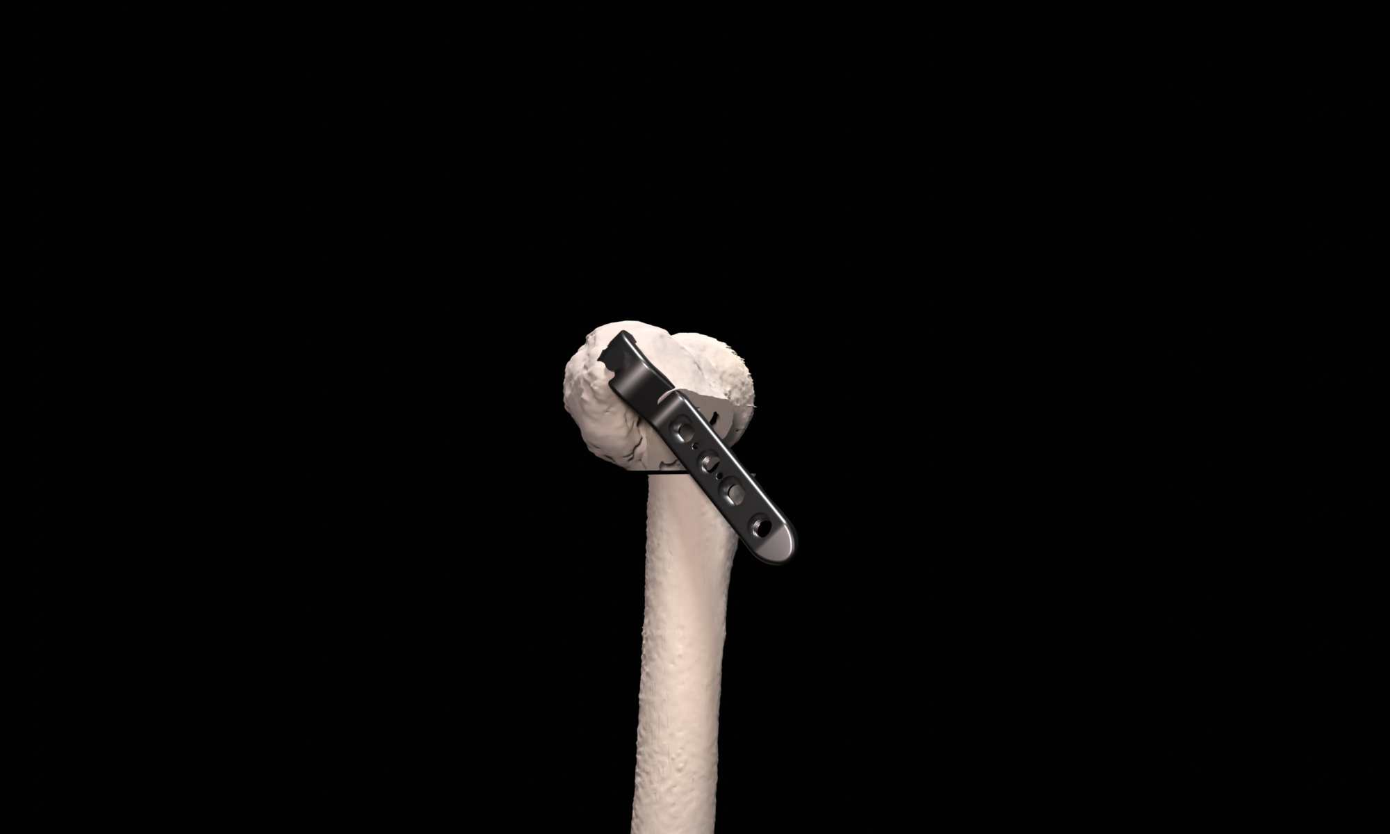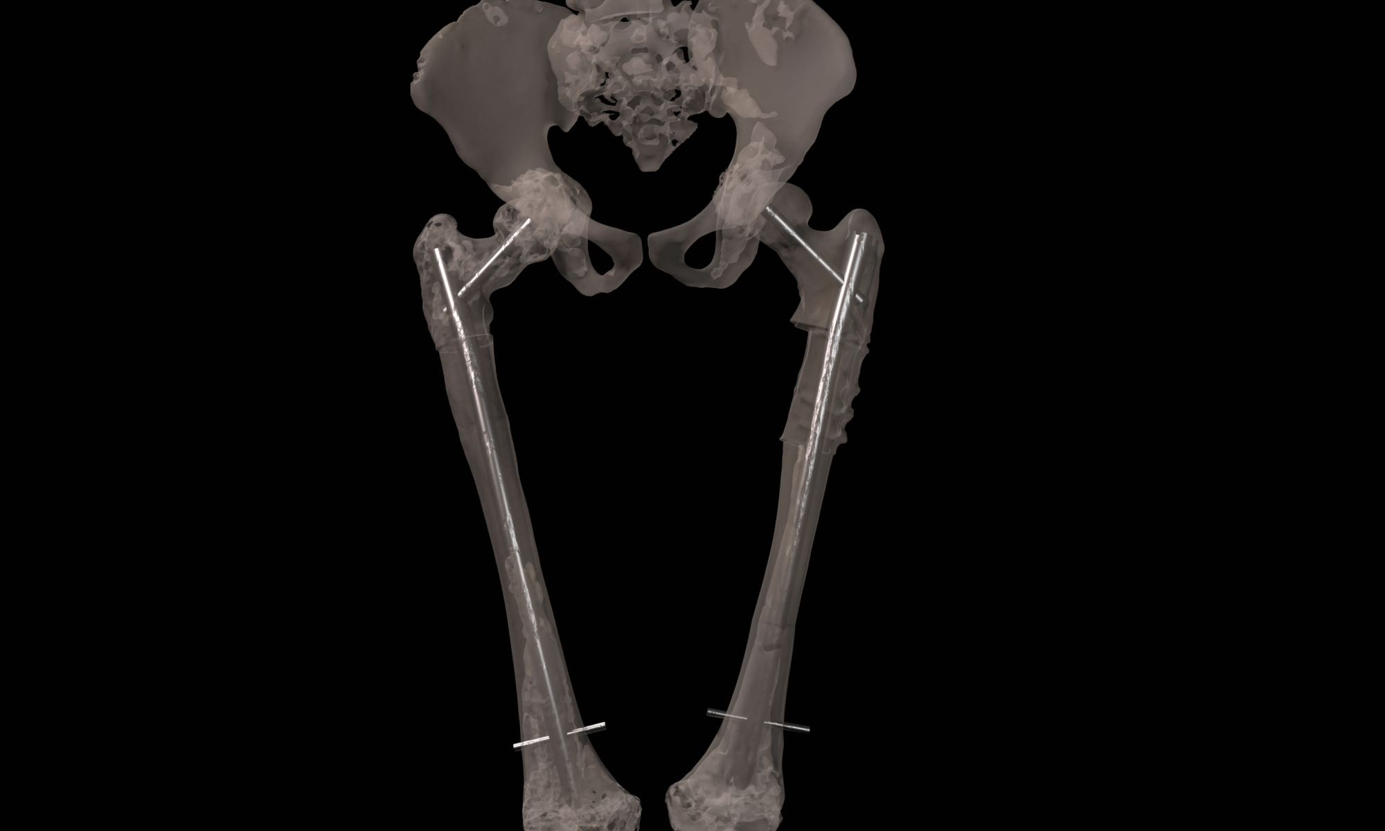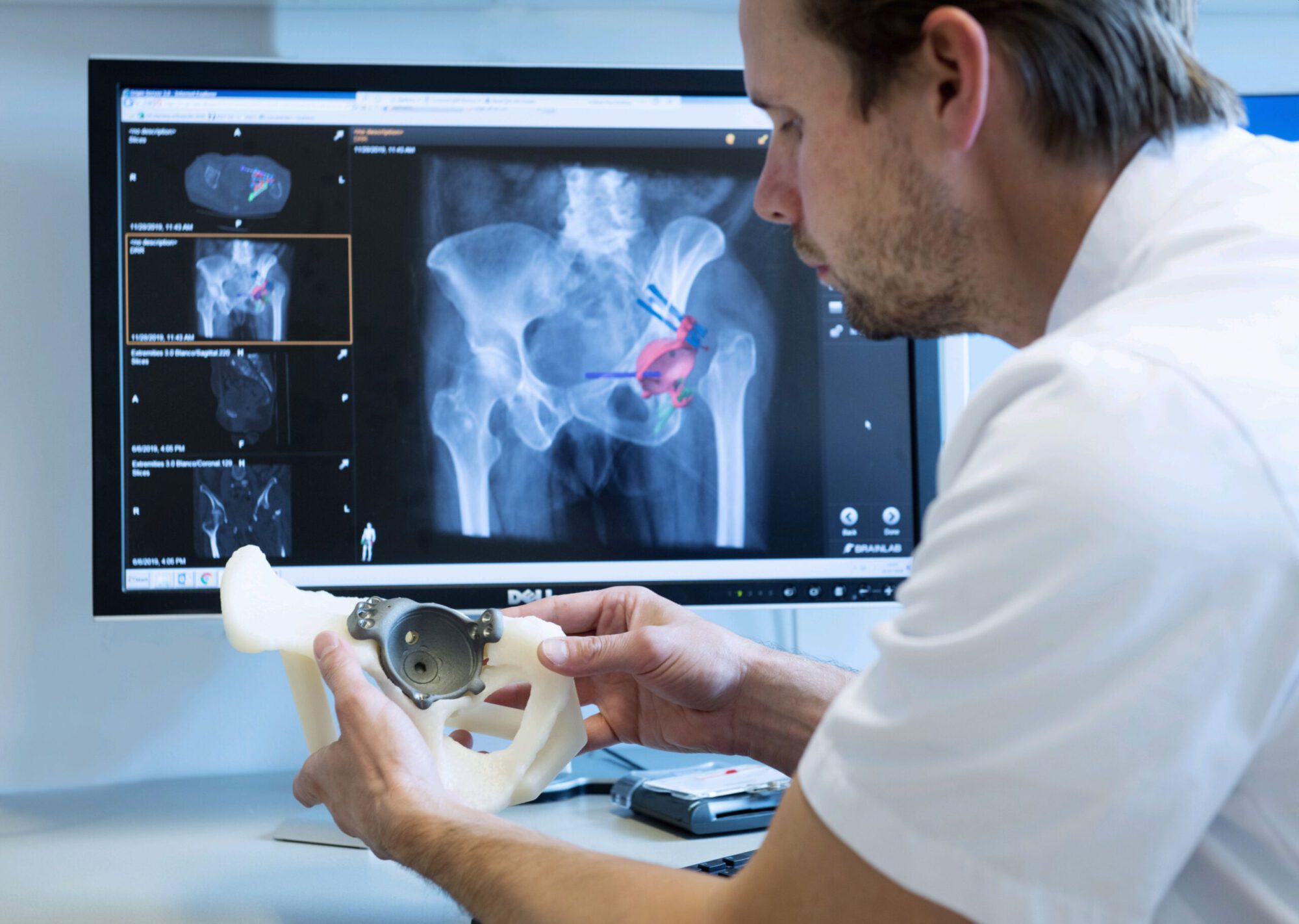
Complex proximal femur deformities can result after (pathological) fracture mal-union. Deformities in this location can be very invalidating due to the relation to the hip joint and negative effect on leg-length, rotations and limb biomechanics. Proximal femoral deformities can be treated by corrective osteotomies and are preferably secured with a compressive blade plate. In complex cases, careful surgery planning in 3D is necessary to achieve the desired correction in multiple planes. A key part of this procedure is to position the blade of the plate in the femoral head and neck correctly. First, the desired post-operative correction and blade position is virtually planned using 3D models generated from 3D CT scans. Second, to translate the virtual planning to the patient in the operating room, patient specific guides are designed, 3D printed and sterilized for intra-operative use. These custom surgical guides, that precisely fit the unique bony shape of the patient, indicate the desired osteotomy location, osteotomy planes and blade direction and facilitate the execution surgical procedure with high accuracy.
Proximal Femur Osteotomy