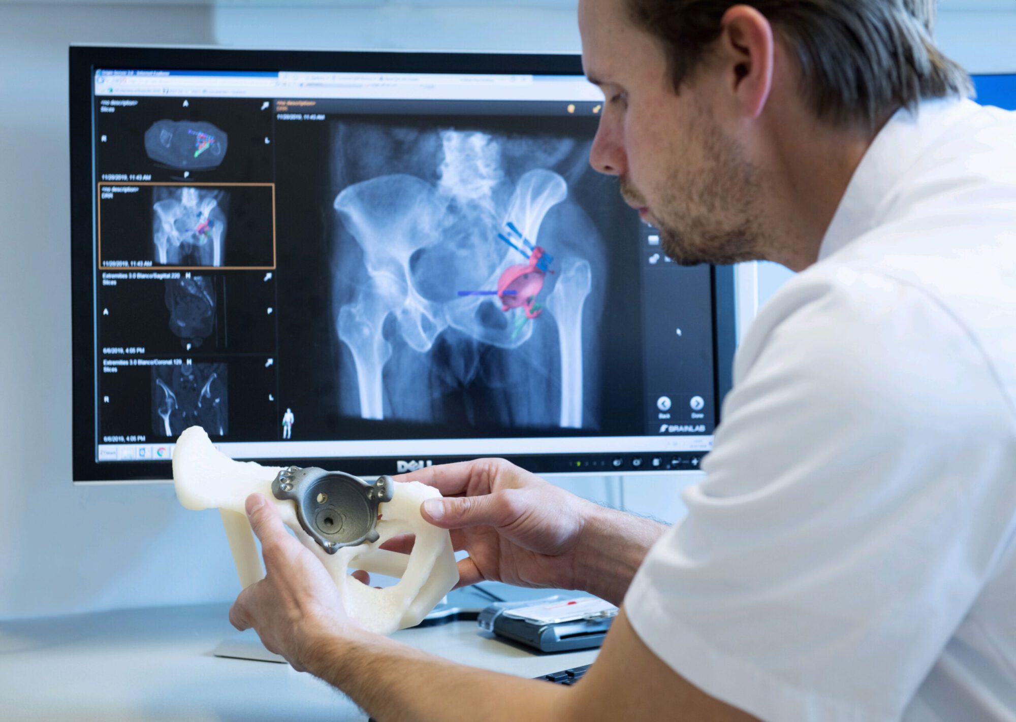
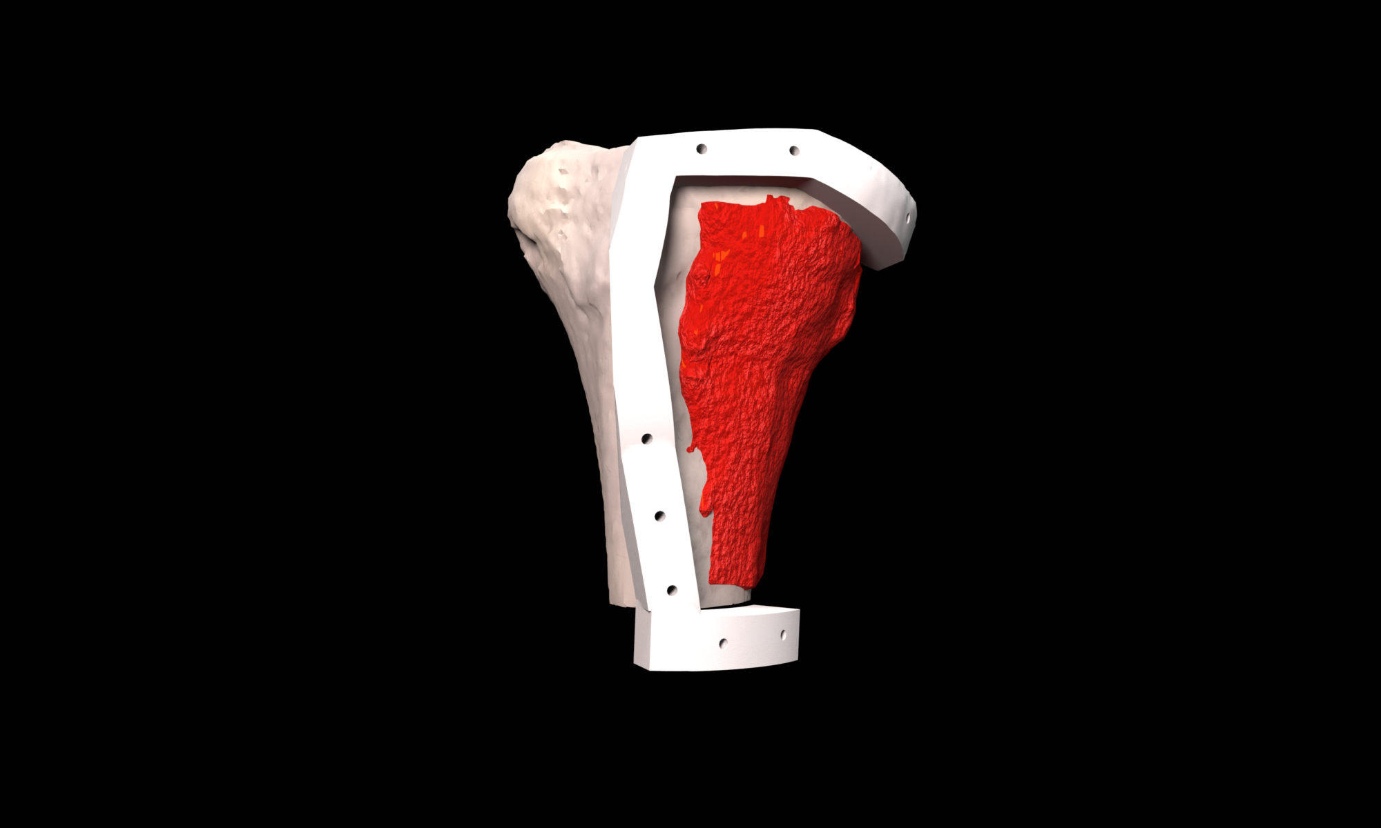
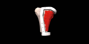
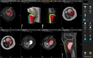
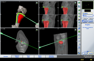
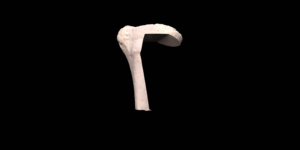
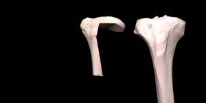
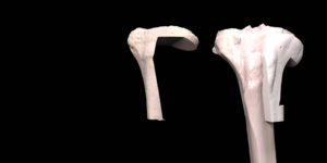
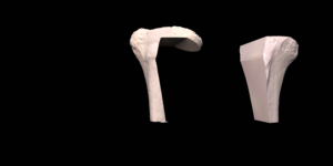
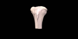
Biological reconstruction is still the preferred method to reconstruct bone defects caused by bone tumor resection in young patients. The removal of the tumor including a required margin is 3D virtually planned in which the information of different imaging modalities, such as CT, MRI and PET, is combined. The surgical resection plan is translated to the operation room by designing patient specific surgical guides that uniquely fit the patient anatomy and indicate the virtually planned resection. To validate the use of these guides surgical navigation is used during surgery. Data from a digital bonebank, that contains 3D models of all available allograft bones, is used to select the best matching allograft bone and to design resection guides for accurate allograft preparation during surgery.
Proximal Femur Osteotomy
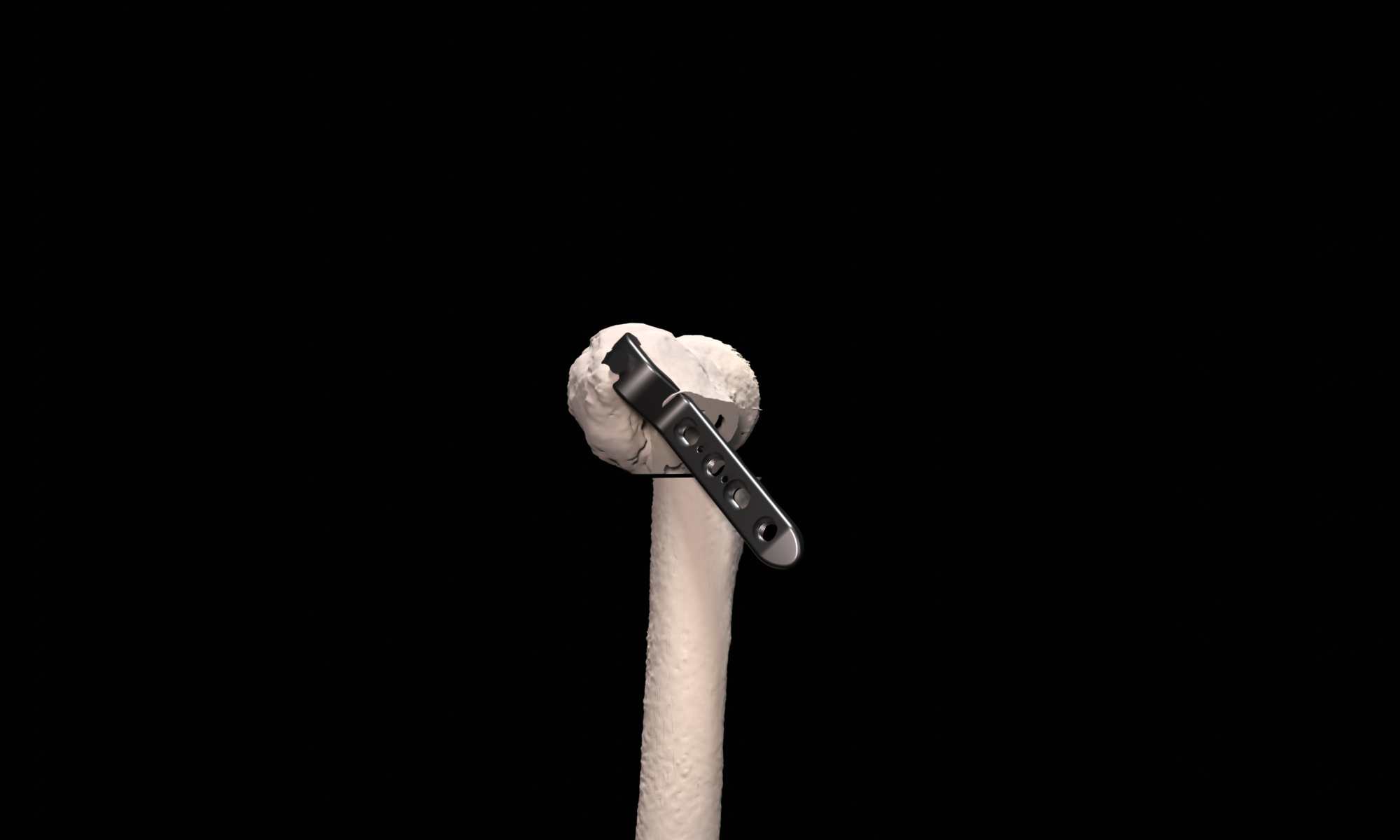
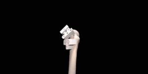
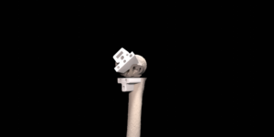
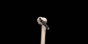
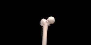
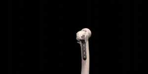
Complex proximal femur deformities can result after (pathological) fracture mal-union. Deformities in this location can be very invalidating due to the relation to the hip joint and negative effect on leg-length, rotations and limb biomechanics. Proximal femoral deformities can be treated by corrective osteotomies and are preferably secured with a compressive blade plate. In complex cases, careful surgery planning in 3D is necessary to achieve the desired correction in multiple planes. A key part of this procedure is to position the blade of the plate in the femoral head and neck correctly. First, the desired post-operative correction and blade position is virtually planned using 3D models generated from 3D CT scans. Second, to translate the virtual planning to the patient in the operating room, patient specific guides are designed, 3D printed and sterilized for intra-operative use. These custom surgical guides, that precisely fit the unique bony shape of the patient, indicate the desired osteotomy location, osteotomy planes and blade direction and facilitate the execution surgical procedure with high accuracy.
Anatomical Models
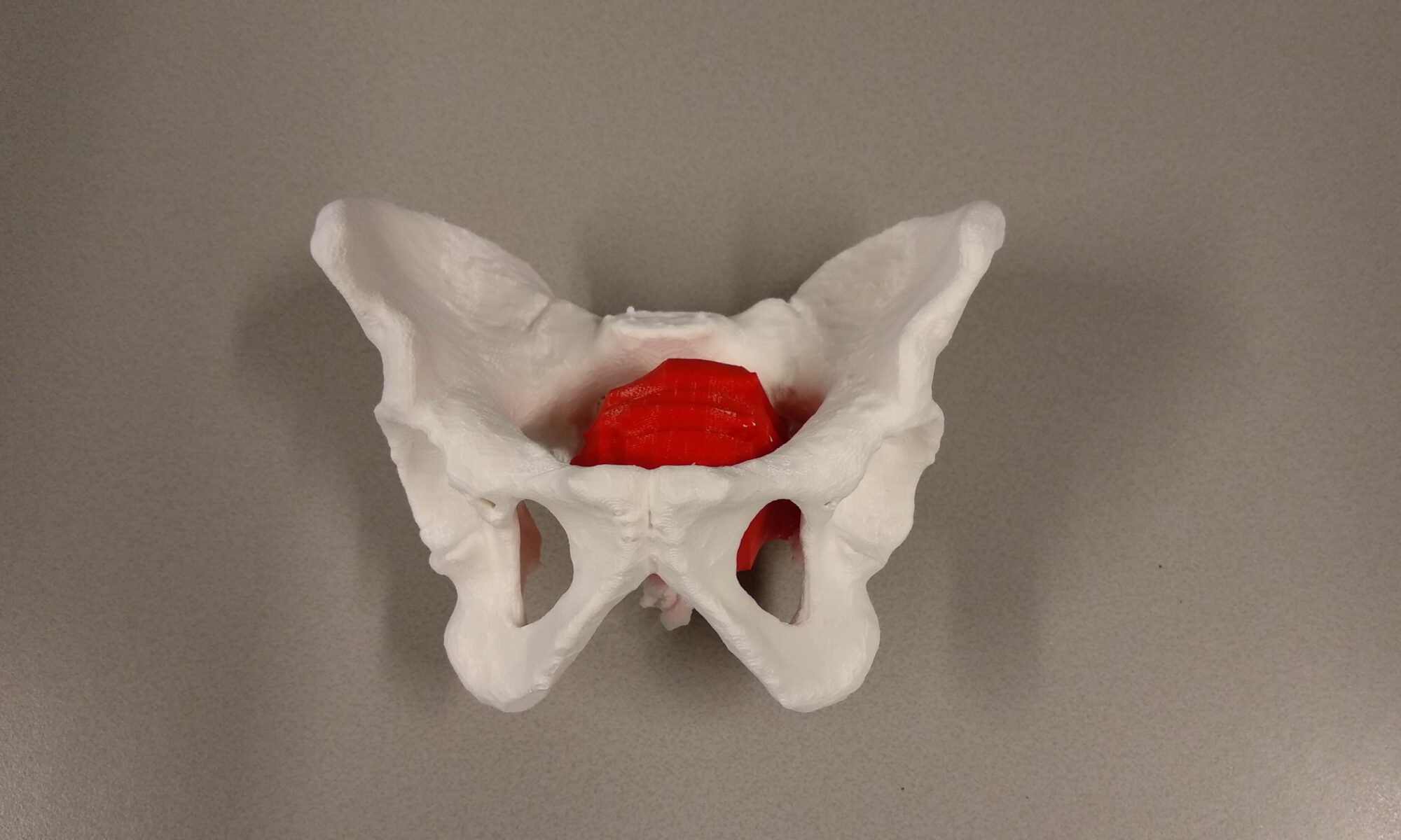
For surgical preparation and shared decision making between multidisciplinary surgical teams, patient specific, multi colored 3D printed anatomical models can be very useful. 3D models of (bony) pathology also have a high educational value for surgeons in training. Also paramount is the use of 3D printed anatomical models for patient insight into their disease and shared decision making between surgeon and patient.
Full Femur Osteotomy
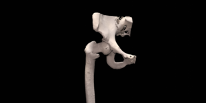
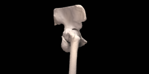
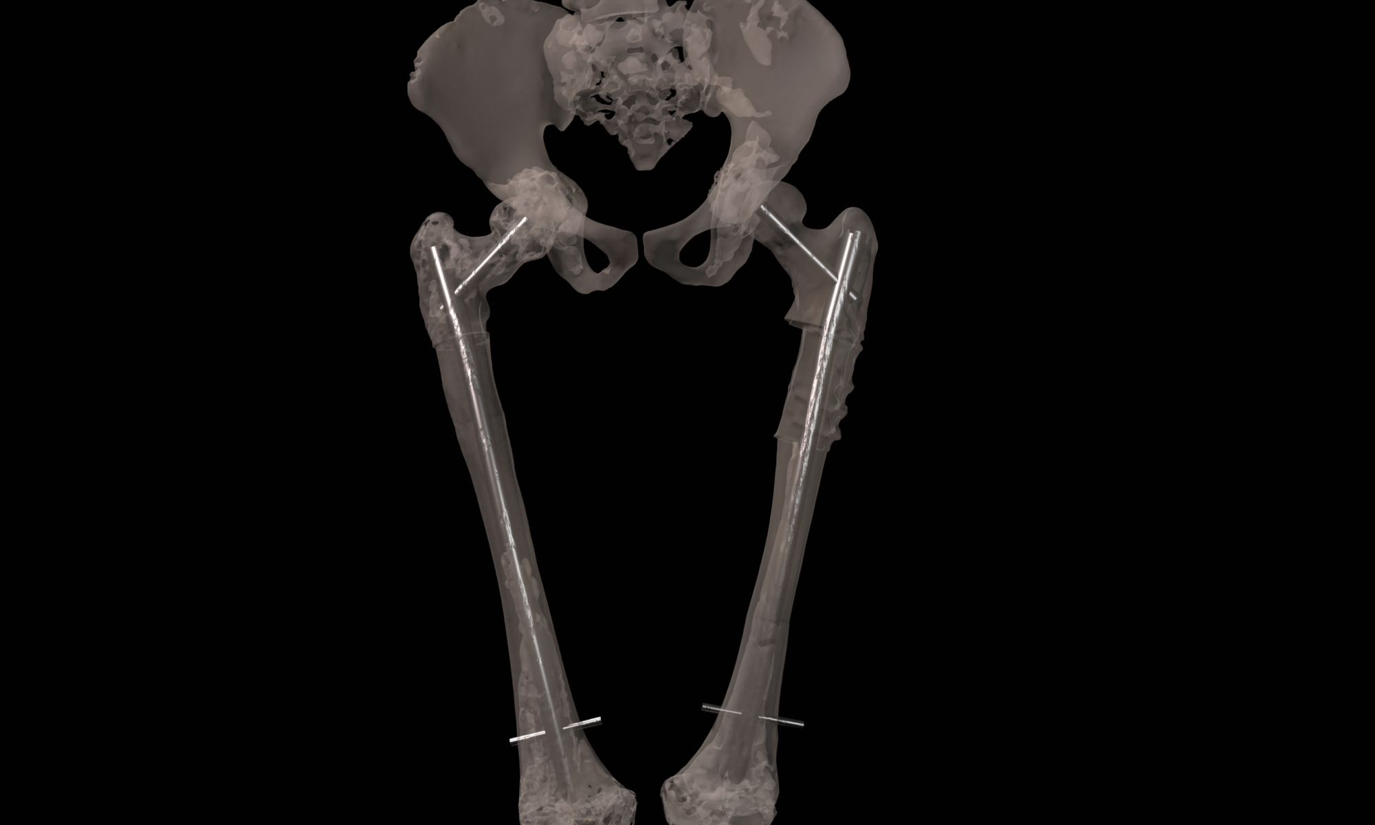
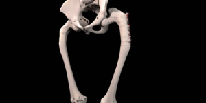
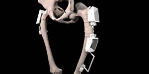
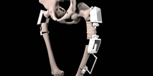
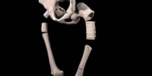
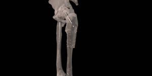
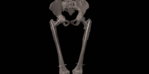
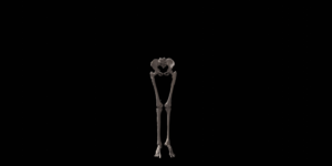
A Shepherd’s Crook deformity is caused by fibrous dysplasia and can be treated by a corrective osteotomy secured with eighter plates or intra-medullary nails. In complex cases, multiple and very large corrections are necessary, requiring careful surgical planning in 3D. First, the desired post-operative correction is virtually planned using 3D models generated from 3D CT scans. Second, to translate the virtual planning to the patient in the operating room, patient specific osteotomy guides are designed, 3D printed and sterilized for intra-operative use. These custom surgical guides, that precisely fit the unique bony shape of the patient, indicate the desired osteotomy location and osteotomy planes and facilitate the execution of the surgical procedure with high accuracy.
The patient in the example case had two surgeries to correct both legs.
